
Ventriculoperitoneal Shunt Placement
What is a Ventriculoperitoneal Shunt (VP shunt)?
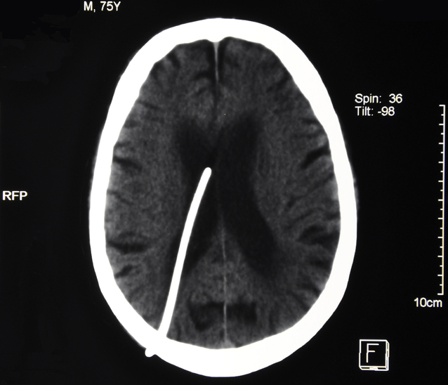
A ventriculoperitoneal (VP) shunt is a device implanted in the brain of a patient with hydrocephalus, which is a buildup of cerebrospinal fluid (CSF) in the brain.
Hydrocephalus is caused by an obstruction that prevents CSF from draining properly. When CSF accumulates, it can enlarge brain ventricles and stretch nerve tissue.
A VP shunt is used to drain CSF away from the brain and into the abdomen, where it is easily absorbed.
Hydrocephalus can develop at any age, but it is more common in babies and older adults.
Hydrocephalus is often present at birth, although it can develop later in life from lesions or tumors within the brain, central-nervous-system infections, or severe head injuries.
Shunts can also be used in conditions such as normal pressure hydrocephalus (NPH) and Pseudotumor Cerebrii (Benign Intracranial hypertension).
The VP Shunt Procedure
Performed with the patient under general anesthesia, VP-shunt placement typically takes 90 minutes. To prepare the area for an incision, a small patch of hair is shaved from the head (usually near the top) or behind an ear.
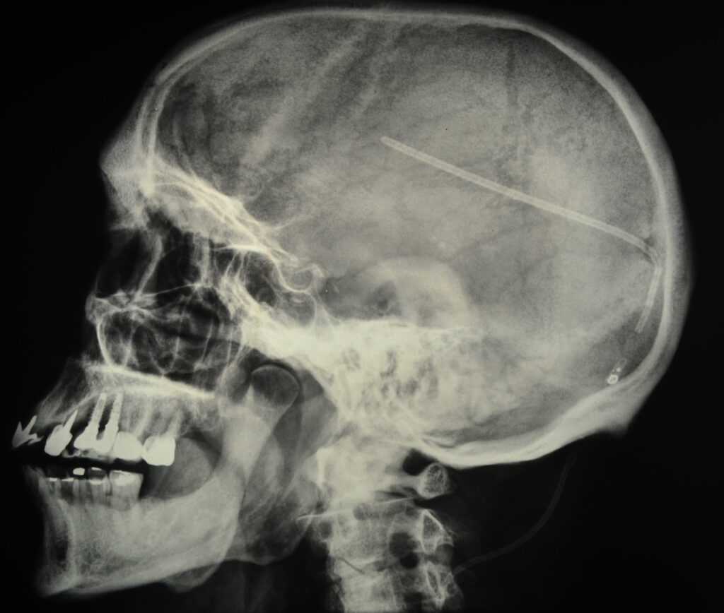
A U-shaped incision is made, and a hole is created in the underlying skull to gain access to the brain. A catheter is then placed within a ventricle of the brain, often with the surgeon using either an endoscope or a computer-guidance system to obtain a clear view of the area.
A second catheter is inserted beneath the surface of the skin toward the back of the head, then threaded down until it reaches the peritoneal cavity of the abdomen. An incision is made in the abdomen to help to properly position the catheter.
Many times a General Surgeon is used to help with the abdominal part of the procedure, placing the distal catheter into the peritoneal space of the abdomen.
A very small pump is also placed at the incision site on the head; it is attached to each of the catheters. As fluid levels begin to increase near the brain, the pump activates, safely draining the necessary amount of CSF into the abdomen.
Many of the pumps used in VP-shunt surgery have programmable or adjustable valves, enabling the flow of CSF out of the brain to be adjusted, which results in more efficient treatment of hydrocephalus.
Learn More About Ventriculoperitoneal Shunts
Risk of VP Shunt Placement
The procedure for placing a VP shunt is considered safe, but it does have some risks. Risks associated with VP-shunt surgery include:
- Shunt Failure or Blockage (obstruction) – is one of the most common problems. Blockages of the catheters can often be fixed (sometimes with further surgery) and rarely result in serious harm.
- Shunt Malfunction – may include over- or under-drainage.
- Over-drainage – When the shunt allows cerebral fluid to drain from the brain’s ventricles more quickly than it is produced, the ventricles can collapse, possible causing a bleed.
- Under-drainage – occurs when CSF is not removed quickly enough. Pressure builds and the symptoms of hydrocephalus recur.
- Infection at the site of the surgical wound, the shunt or in the cerebrospinal fluid itself (meningitis). Symptoms may include a low-grade fever, soreness of the neck or shoulder muscles, and redness or tenderness along the path of the shunt. Hydrocephalus symptoms may reappear as well.
- Brain bleed
- Abdominal complications such as bowel perforation, bowel obstruction, bowel abscess, abdominal cyst formation.
Schedule an appointment
- Contact Us
Ready to learn more? Contact us today to get started.
Make an Appointment
New & Existing Patients: Request An Appointment
New and existing patients may request an appointment by filling out the form below. Someone from our team will be contacting you to schedule an appointment time.